Showing posts with label dean toriumi review. Show all posts

Tuesday, December 3, 2013

Changes Made to My Technique Over the Past Six Years

Over the past 6 years several changes have been made to improve our techniques. These changes were made in an effort to improve our outcomes and improve patient satisfaction.

One of the biggest changes is the way I am managing the nasal tip cartilages. Over the past six years I have moved to repositioning the lower lateral cartilages into a more favorable position. This concept is complex and may be difficult to understand however I will try to explain it. Many patients that have a bulbous nasal tip exhibit cartilage anatomy that creates the roundness seen on frontal view. (Figure 1)

Figure 1
This bulbous shape is frequently created by the patient’s cartilages being large and round bulging outward to create the poorly defined shape. Another contributing factor is the angulation of the lower lateral cartilages as they move cephalically or upward.

In the ideal nasal tip there is less bulk in the supratip area of the nose (area just above the nasal tip as shown in this illustration that was developed from a composite of many ideal nasal tips and merged into a single “ideal” nasal tip. (Figure 2)

Figure 2
This is not universal but has many of the favorable characteristics of an aesthetically pleasing nasal tip. In our review of patients with aesthetically pleasing nasal tips we also found that they possessed either flat lateral crura (tip cartilages) or have lateral crura that move in a caudal orientation. Caudally oriented lateral crura move laterally at a larger angle (greater than 30 degrees of off midline). Most bulbous tips will have lateral crura that move at a more acute angle off of midline (less than 30 degrees). This cartilage orientation is referred to as “cephalically positioned lateral crura.” These angles are averages and not absolute. There are some patients that have an angle less than 30 degrees and still have nice nasal tips but most of these patients will also have relatively flat cartilages. Most patients with an angle less than 30 degrees and convex cartilages will have a bulbous nasal tip contour.

To better understand this concept one can think of the nasal tip as a series of shadows and highlights. Shadows are created by concavities and highlights are created by convexities. If we look at the actual nasal tip cartilage anatomy in a patient with a bulbous cephalically positioned lateral crura the tip cartilages pass directly through the area where the shadow of the supratip should be located.(Figure 3)

Figure 3
If the tip cartilages are moved to a more caudal orientation (angle greater than 35 degrees) the area of the supratip is cleared of the convex cartilage and a shadow is created.(Figure 4)
Figure 4

Additionally, an area of highlight is created along the rim of the nostril near the base of the nose similar to the artist’s rendition of the ideal nasal tip. It may be difficult to understand that increased width or bulk is good in some areas of the nasal tip but this is exactly what we see in the favorable nasal tip. We see areas of shadow in the supratip and lateral supratip with areas of highlight along the margin of the ala. This area of highlight along the margin of the ala is represented by a uninterrupted triangular shape of the base of the nose. (Figure 5)
Figure 5
In the pinched nasal tip there tends to be a shadow just lateral to the tip which acts to isolate the tip making it appear less desirable and in some cases more bulbous.(Figure 6) Examination of the base view of such a nasal tip will usually show an interruption in the desired triangular shape to the base view. (Figure 6)

Figure 6
In the pinched tip the shadows extend into areas that are not favorable and these shadows are due to concavities or depressions just lateral to the tip. Many commonly used nasal tip techniques tend to narrow the nasal tip cartilage without considering that good support should be preserved along the caudal margin of the lateral crura and to preserve or create a triangular shape to the nasal base. Again it is counterintuitive that one should believe that areas of the lower lateral cartilages should be left prominent as most would think that all around smaller would be better. Unfortunately, that is not the case. In order to create proper shape the skin envelope must be controlled creating shadows and highlights, preferably favorably positioned shadows and highlights. Therefore, creation of an aesthetically normal looking nasal tip is not necessarily narrow and a narrow nasal tip is not necessarily aesthetically normal. Nasal tip surgery is complex and far more complicated than simply an exercise in suturing the tip cartilages or placing a tip graft.

Patients that have cephalically positioned lateral crura will also tend to have weakness in the lateral wall of their nose. This weakness can contribute to collapse of the lateral wall of the nose. This type of collapse can be easily seen on base view as the patient breathes in through their nose (Figure 7).
Figure 7
If one side collapses and the other does not it is likely due to a significant septal deviation in addition to the lateral wall weakness. This is because most of the nasal airflow is passing through the side opposite the septal deviation. This airflow overwhelms the lateral nasal sidewall on the side opposite the deviation and the flow of air collapses the nostril on that side. In order to correct collapse of the nasal sidewall the first step is to correct the septal deviation to create more symmetric airflow through both nostrils. Airflow through both nostrils is rarely symmetric but the key is to eliminate significant differences in airflow.

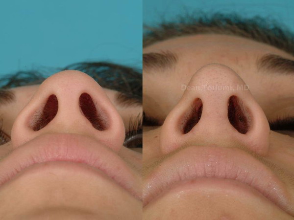
Correction may also require some conservative work on the inferior turbinate which can be enlarged on the side opposite the septal deviation. Once the internal nasal airway problems are corrected weakness of the lateral wall of the nose should be addressed. If not corrected the patient may be left with a compromised nasal airway. Correction of lateral weakness can be accomplished using several different methods. One of the more common methods is to insert an alar batten. These grafts are placed into the sidewall of the nose and provide extra support to help prevent lateral wall collapse. These grafts can work well but typically do not improve nasal tip shape. In order to improve nasal tip shape I frequently “reposition” or move the lateral crura into a more caudal orientation. In this technique the cephalically positioned lateral crura are dissected from the underlying vestibular skin and then moved into a more favorable orientation or more obtuse angulation off of midline (greater than 35 degrees in most cases). This technique is not new and is used by other surgeons as well.  Since increasing the use of lower lateral cartilage repositioning I have found two major benefits to the patients. First of all, these patients have their lateral crura moved to a more caudal orientation which acts to support the lateral wall of the nose, minimize lateral wall collapse and improve nasal airflow. The reason for this improvement in nasal function is that the cartilage is moved to an area where the lateral wall has a propensity to collapse and provides support that helps to prevent collapse. (Figure 8 A,B and C)


The other major benefit is that the tip cartilages are moved into a position that improves nasal tip shape by moving bulk from the area of the supratip (area above nasal tip) to the area of the inferior tip. In the ideal nasal tip there should be a gentle widening of the nose as the lateral wall approaches the inferior nostril margin.
Postop frontal view
This favorable shape creates the normal nasal contour seen in the artists composite from our study of aesthetically pleasing nasal tips. If the nasal tip is pinched and the normal flare toward the nostril rim is not present the nose may look unnatural (Preoperative view).

Preoperative view
This “pinched” look gives the nose an operated look and can be associated with poor nasal function. The one drawback is that this maneuver creates increased stiffness to the lateral wall of the nose and nasal tip. It is this increased stiffness that improves nasal function. In most patients this is an acceptable trade off. If there is little support in the lateral wall of the nose collapse is likely to occur at some point in time. All too often patients have little concern about their nasal function when they undergo rhinoplasty and just want their nose to look good. Unfortunately, if the nose is weakened and there is inadequate lateral wall support nasal function may become compromised over time. This will likely catch up with most patients later in life. A more sensible approach is to provide good lateral wall support and enhance aesthetics with the tradeoff of some increased stiffness in the sidewall to provide good long term breathing. (Figure 11)


In order to reposition the nasal tip cartilage a cartilage graft (lateral crural strut graft) is sutured onto the undersurface of the lateral crus and then the cartilage is placed into a newly created pocket in the sidewall of the nose. It is this additional layer of cartilage that creates the increased strength and stiffness but also provides the improved support. Placing lateral crural strut grafts requires two relatively large pieces of cartilage. Usually these cartilage grafts are harvested from the nasal septum.

I also use spreader grafts in most patients which are rectangular shaped grafts that act to reconstruct the middle portion of the nose below the nasal bones. These cartilage grafts act to prevent middle nasal vault collapse or the “inverted-V deformity.” (Figure 12)

Figure 12
The inverted-V deformity indicates that the cartilages in the middle portion of the nose have collapsed and also can compromise nasal breathing and create deformity. Spreader grafts are typically used after dorsal hump reduction to recreate a stable “roof” to the middle segment of the nose.(Figure 13)


 The other common cartilage graft that I use is the caudal septal extension graft. This is a cartilage graft that is typically placed as an extension off of the inferior part of the septum. This graft preserves nasal tip projection and helps to prevent the “polybeak deformity” by preventing postoperative loss of nasal tip projection . The caudal extension graft is similar to a columellar strut but is fixated to the patients existing nasal septum to provide additional support.(Figure 14)

Figure 14
This additional support stabilizes the base of the nose to increase and support nasal tip projection.(Figure 15)


The caudal septal extension graft also adds some stiffness to the nasal tip. This is a reasonable trade off to provide a stable nasal tip that will not lose projection postoperatively.  

The techniques described require a significant amount of septal cartilage and in the past I have gone to the ear or rib to have enough cartilage to execute these grafting maneuvers. Another new addition to my technique is the use of a resorbable (dissolvable) implant made from a suture material that can be used to stabilize caudal septal extension grafts. In the past I would need to make spreader grafts long enough to support the septal extension grafts. With the use of the resorbable “PDS” plates I can stabilize the extension grafts without using extra cartilage for that purpose. (Figure 16)


The PDS plates a completely gone from the patient in about 6 months.
I am also able to occasionally join two pieces of cartilage together to make a single longer graft that can be used for a lateral crural strut graft or spreader graft. The incorporation of the PDS plate has significantly decreased the need to harvest cartilage from the ear or rib. In fact, I rarely harvest cartilage from the ear or rib in primary rhinoplasty patients. The primary rhinoplasty patients that require rib cartilage are typically undergoing complex reconstruction or major augmentation such as saddle nose deformity, major dorsal nasal augmentation in Asian patients, severe crooked nose deformities or congenital nasal deformities. These are complex cases requiring a large amount of grafting material to correct their deformities and rib cartilage is abundant and strong for a stable reconstruction. 

If I harvest rib cartilage I use a very small 1.0 cm to 1.1 cm incision placed just below the right breast. (Figure 17)
Figure 17
This incision usually gets stretched to 1.3 cm. This small incision looks like a small stab incision once it is healed. In the past I would make a larger incision that was more prominent and was noticeable to patients. This small incision is rarely an issue with patients as it usually barely visible once the redness fades. (Figure 18). 
Figure 18
This small incision is important to most patients as the presence of a large chest scar on a patient’s body will be a prominent visible reminder of their surgery. 

Another change that I have made is that when I bank cartilage I only place smaller softer pieces in a small incision behind the hairline. These cartilage grafts are ideal for filling small defects in the nose or providing a subtle contour change. The banked cartilage is very helpful in cases where a revision is needed as the ear or rib is rarely needed. In patients that are more likely to need a significant revision I prefer to bank the larger pieces of cartilage that may be bothersome to the patient but can be removed at a later date.


Friday, August 30, 2013

Dean Toriumi: Facelift Surgery

If you are unhappy with the lower two-thirds of your face, Dr. Toriumi may suggest you undergo a rhytidectomy procedure. A rhytidectomy, or facelift, is designed to correct conditions associated with an aging face. Visible indicators include saggy or loose skin and loss of facial muscle tone. A facelift can correct these indicators by removing excess skin from the face and neck, and sculpting the underlying tissues and muscles in your jaw, cheek, and neck area.
A facelift can be combined with other procedures to strengthen your chin and improve your brow or eye area. These include brow, chin, or eyelid surgery. 

Pre-Operative Instructions: Before Surgery:

This information is designed to answer questions about your pre-operative care. To achieve the most satisfactory results, you should follow these instructions closely and familiarize yourself with these instructions. Please attempt to follow these instructions faithfully.

1. Do not take any aspirin, compounds containing aspirin, or non-steroidal anti-inflammatory medications (Motrin, Ibuprofen, Advil, Aleve, Vioxx, Celebrex, Naprosyn, etc.) for four weeks prior to and four weeks after your surgery.
2. If you require pain medications, you may safely take Tylenol products in the period from surgery preparation until you return home. If you require stronger pain management, consult Dr. Thomas.
3. Do not take Vitamin E or herbal supplements such as gingko, ginseng, fish oil, garlic, St. John's wort, etc. for four weeks prior to your surgery and four weeks after your surgery.
4. Avoid alcoholic beverages 48 hours prior to surgery.
5. If you smoke tobacco or use smokeless tobacco products, stop using them at least four weeks prior to surgery and refrain from them for at least four weeks after surgery. Smoking is directly related to poor wound healing and possible skin loss.

Post Surgery:

To expedite healing and minimize discomfort, please follow Dr. Toriumi's explicit directions. Dr. Toriumi will advise you to avoid strenuous activities right after surgery.


Wednesday, August 21, 2013

Dean Toriumi: Anti-inflammatory Diet

Dean Toriumi: Anti-inflammatory Diet


Many prominent physicians –including Dr. Barry Sears, Andrew Weil, and “Dr. Oz” – and dozens of books have advocated healthier, anti-inflammatory diets for improved health and weight loss. A few have addressed the connection between diets and allergy. But Dean M. Toriumi, M.D., a noted facial plastic surgeon and Professor of Otolaryngology-Head and Neck Surgery at UIC, has adapted the antiinflammatory diet trend to his facial plastic surgery patients – with significant results, results that help improve healing and quality of life for many patients post-surgery.

Many Americans consume diets high in pro-inflammatory foods – processed fats and meats, hydrogenated oils, trans fats, and refined grains and sugars. These ingredients can radically affect how we respond to food ingredients that can induce allergic reactions, often instantly. On average, the more inflammatory substances we put into our body, Dr. Toriumi noted, the greater the allergic response. Symptoms - from congestion and sneezing to watery eyes and skin irritation - can result. For Dr. Toriumi, a world-renowned facial plastic surgeon, the effects of inflammation on his patients’ nasal symptoms, especially among those recovering from surgery, were of particular concern. Nasal congestion, breathing difficulties, and related inflammation problems can be amplified in surgical patients, causing significant discomfort and slowing recovery –an important factor for patient outcomes.

So Dr. Toriumi began prescribing his anti-inflammatory diet – developed over many years and has some similarities to the Sears “Zone Diet” and the proven Mediterranean diet – to surgical patients. “Some facial plastic surgery patients have expectations about how quickly they’ll get the look they’re seeking from surgery,” he noted. “While some patients clearly have unrealistic expectations that everything will look great instantaneously, clearly diet can make a difference, expediting people’s recovery by contributing to a reduction of swelling,” he noted.

Dr. Toriumi’s clinical studies found that the diet, when adhered to in a highly disciplined way, contributed to many patients’ health and recovery. He would prescribe his diet – including a lot of leafy green and vegetables, minimal animal fat (unless it’s from grass-fed animals who don’t consume a lot of corn – corn ingested either directly through their body or through an animal that ate corn is highly inflammatory, he noted), green tea, and olive/fish oils – to patients for three months and asked them to record their symptoms. “Among those who were really disciplined and committed to the diet, the results were astounding,” Dr. Toriumi noted. “Regardless of their nasal problem before the antiinflammatory diet – congestion, swelling, breathing difficulties, etc. – the symptoms were significantly reduced and quality of life improved demonstrably with the diet,” he said.

Patients felt better, noticed speedier recovery from surgery, and were better able based on the symptoms they had to understand and gauge those foods that particularly contributed to their allergic conditions. From there, Dr. Toriumi noted, they were better able to dictate and control their own intake of the most inflammatory foods to help manage their diet and allergies going forward, he said. And there were other benefits – aside from the weight loss that typically occurs when people adopt an antiinflammatory diet: patients needed fewer or no medications to manage their allergies. “Because the body through food and nutrition is naturally preventing allergic reactions, many patients don’t need any more pharmaceuticals to do the job for them,” Dr. Toriumi said.

His focus on nasal symptoms, diet, and how it these relate to surgical patients applies to patients both pre- and post-surgery, even years after their initial surgery. In short, with the right diet, anything inflammatory gets better. This is the case for any allergy, he added, whether food-based or not. “It all has to do with the body’s response to the allergen and its connection to anti-inflammatory agents,” he noted.

As a facial plastic surgeon, Dr. Toriumi’s work is unique among the tomes of diet books and TV shows we see today. “It is a bit out of left field for a surgeon to look into these issues, but it feeds well into my practice and patients,” he noted.

“Most patients really want to take care of themselves and be in control of their health. Particularly among surgical patients, who tend to be well educated and sophisticated – not to mention very much in tune with any nasal symptom that gets in the way of their recovery and outcomes, this is right up their alley,” Dr. Toriumi noted.

Tuesday, August 20, 2013

Dean Toriumi Gives You Balanced Features

Dr. Toriumi Gives You Balanced Features


This patient presented with an under-projected chin.
To help balance her nose with her other facial features, Dr. Toriumi augmented her chin and increased the projection of her her nasal tip.

The photographs represent a two year postoperative outcome.
Pre-Operative PhotosPost-Operative Photos

Thursday, August 8, 2013

Dean Toriumi: Secondary Rhinoplasty surgeries

Secondary Rhinoplasty: 3

Why does Dr. Toriumi only use rib cartilage for secondary rhinoplasty surgeries? 

Dean Toriumi's Answer: 

I noted that when I used ear cartilage for dorsal augmentation, the edges of the ear cartilage grafts would tend to curl or deform over time leaving dorsal irregularity. I also noted that if I stacked multiple layers of auricular cartilage to gain height on the nasal dorsum, some of this cartilage resorbed over time. For this reason I usually avoid stacking more than two layers of ear cartilage. Less severe deformities such as those limited to the tip, lateral wall of the nose or less severe middle vault collapse often can be corrected with ear and/or septal cartilage.


It has become apparent to me that most patients who come to my office are prepared to hear that I may need to use a rib graft in their surgery. It is true that most secondary rhinoplasty patients will need a rib graft to get the maximal outcome. However, on occasion, a patient's rib cartilage is calcified or they prefer that I use ear cartilage instead of rib cartilage. In many of these patients I am able to do a more than adequate reconstruction using ear cartilage. I actually have more experience using ear cartilage for secondary rhinoplasty than I do with rib cartilage. It has only been over the past six years that I have gone primarily to rib cartilage for secondary rhinoplasty, although I have used rib cartilage for over 18 years, but, in my early years in practice, I only used rib cartilage in more severe cases that required a lot of grafting material or when both ears were already taken. As I gained more experience with rib grafting we found that the patients did well and the pain from the rib graft harvest was no greater or less than the ear cartilage harvest. For many surgeons, harvesting and using rib cartilage is a big ordeal with a large incision, risk of a collapsed lung, and bending or warping of the cartilage. Over the years I have become very efficient harvesting and using rib cartilage leaving the patient with only a very small scar (usually less than 2 cm), short-lived postoperative pain, and very low risk of bending or warping of the rib cartilage.


Over the past three years I have done more than 300 rib grafts in patients and have further refined my technique so I am able to make the grafts much thinner and smaller without warping. We measure the thickness of nearly all structural grafts placed in the nose. Using these measurements I know what graft thickness is necessary for a narrow nose or wider nose. This allows me to reliably control the width, length and rotation of the nose. Unfortunately, numerous sources have deemed that I like "big wide noses." I can assure you that this is not the case. It is true that many patients will have swelling early postoperatively that will make their nose wider and larger. However, with time, this swelling will dissipate, and the nose will look narrower and smaller. Patients with thick skin are at the highest risk of being swollen for a long period of time. With healing almost all noses will get smaller and narrower over time. Therefore, our patients will improve over time and tend not to collapse, as I have seen in patients who had ear cartilage grafts placed in surgery. The postoperative follow-up is critical to insuring that the nose heals properly. Some patients will need to perform nasal exercises or tape their nose to create the proper width and shape. If patients do not come back for their follow-up they are at much higher risk for a suboptimal outcome.


If a patient desires to have ear cartilage used for reconstruction and the defect is amenable, I am willing to consider using ear cartilage as long as he or she understands the differences in the potential outcome. The disadvantages with ear cartilage are that the long-term outcome may not be as good with ear cartilage compared to rib cartilage, with a higher chance of unfavorable changes occurring years after surgery. This problem with ear cartilage is apparent in many patients who have undergone a revision every two to five years after using ear cartilage. The nose initially looks good and then narrows and pinches over time. When performing reconstructions on patients with ear cartilage in place, the ear cartilage grafts are very weak and are frequently deformed. The ear cartilage frequently breaks apart when it is dissected and is usually not usable. Because of the lack of ear cartilage strength, it is useful as a filler graft but not as a structural graft. There also may be some change to the shape of the ear donor site and the initial postoperative pain with the ear cartilage is frequently greater than harvesting rib cartilage.


With rib cartilage the patient will have a scar at the bottom of the right breast. In most patients this scar heals nicely. We have decreased the length of our rib cartilage harvest scars to 2 cm or less. Most of our scars measure between 1.5 cm to 1.7 cm . This smaller scar is much easier to hide. Patients in whom I use rib cartilage for grafting usually will note that they have a stiffer nose, which can be worrisome to the patient, but improves over time. The more dramatic the deformity, the more likely the nose will be stiff because of the need for more structural grafting in the severely deformed nose. We have made our columellar struts thinner and more pliable, which makes the nose less stiff with a more normal feel. I think these refinements have allowed me to make much smaller and narrower noses. Rib cartilage grafting is very technique-dependant and executing these techniques requires a great deal of experience. This is a very important advancement in rib grafting and will allow patients who desire smaller noses to be treated with rib cartilage. 


image


image


image


image


image


image


I felt compelled to answer this question on my website because I have been told by many patients that those interested in secondary rhinoplasty who do not want a rib graft do not come to see me. I was also told by many patients that other surgeons tell them that Dr. Toriumi only does rib grafts. I can assure you that my experience is not limited to rib grafting, and I can always call on my years of experience using ear cartilage to correct secondary rhinoplasty deformities in patients with deformities that are treatable with ear cartilage. It is also important to know that I will not compromise a potential outcome to use ear cartilage in a patient who really needs a large amount of cartilage to correct their deformity.


The primary reason that I have transitioned to rib cartilage is that I was frequently frustrated by the scenario where I was short on cartilage and forced to make a compromise on certain grafts during surgery. By following these patients long term I found that the compromised graft frequently resulted in an unfavorable outcome many years later. Now I am rarely in this situation because there is always enough of rib cartilage available. Seven years ago it was a big deal to take a rib graft with longer operating time and longer recovery. At this time harvesting rib does not add much time to the surgery and it has become routine. In my hands the disadvantages to using rib cartilage are few but the advantages are numerous. 

Tuesday, August 6, 2013

Surgeon’s Review of Dr. Dean Toriumi’s Surgical Techniques

Personal experience visiting Dr. Toriumi   
- Jochen Wurm, M.D.    


During the last 3 years for two weeks at a time I had the great opportunity and privilege to visit Dr. Toriumi in his operating room and observe his surgery. From the very beginning he created a very warm and welcoming atmosphere for me as a guest from Germany. Also his fellows and his staff supported me with great efforts to get familiar with the environment and the routine work flow.My primary intention to visit Dr. Toriumi was to learn more details about his renowned surgical techniques I had watched so many times during various rhinoplasty meetings around the world. What Dr. Toriumi then offered me at his office exceeded all my expectations by far. Prior to the actual surgery he already shared his thoughts on how to prepare the patient and explained his surgical plan in detail. During surgery he paid very close attention to me being able to follow every single step of his work. Dr. Toriumi took additional time to make drawings for my better understanding. I also experienced his extensive efforts to illustrate all surgical techniques in a way that was easily understandable for me in order to overcome any possible language barrier. I was welcome to ask questions at any time and Dr. Toriumi was always willing to answer them in detail. I never had the impression he kept any secrets from me, he rather gave me the opportunity to get profound insight into his broad fund of tips and tricks in rhinoplasty.

I never had the chance to observe a surgeon who performed rhinoplasties with such patience and accuracy as Dr. Toriumi. He took care of any palpable or hardly even visible irregularity and asymmetry that most surgeons would not even recognize. He never finishes surgery until he achieved the best possible result for the patient – whatever it may take, even in the most difficult cases. As a colleague in facial plastic surgery I can only highly appreciate Dr. Toriumi´s outstanding and long-lasting results in rhinoplasty and facial plastic surgery in general.

Both professionally and personally being with Dr. Toriumi was a unique learning experience for me. His inspiring way of teaching enabled me to apply his advanced surgical techniques and attitude on my own patients with great success.

I am very grateful to have met Dr. Toriumi in person. I sincerely would like to thank him for giving me the opportunity to observe his surgery, last but not least also for his kindness and patience.

Therefore I am looking very much forward to meeting Dr. Toriumi again in the near future


Erlangen, Germany
July 24, 2013
Jochen Wurm, M.D.

Friday, August 2, 2013

Dr. Toriumi's operation to correct the crooked nose deformity

Why does Dr. Toriumi use such a complex operation to correct the crooked nose deformity? 

Straightening crooked noses is one of the most difficult tasks in rhinoplasty. Crooked noses can occur for many different reasons. These include prior trauma or asymmetric nasal growth. Deformities can be broken down into two major categories. These include bony deviations (upper half of the nose), and cartilage deviations (lower half of the nose). Bony deviations can frequently be corrected with osteotomies or creating controlled fractures in the nasal bones and shifting them back to the midline. Deviations of the cartilaginous part of the nose (lower half of the nose) tend to more complex and more difficult to correct. Many of the deviations of the lower third of the nose involve deviations of the underlying nasal septum. In some cases less complex surgeries can correct deviations of the lower half of the nose. However, the incidence of failure is higher when an operation does not successfully straighten a deviated septum. Nasal function is also maximized by creating a straight nasal septum. My intent with deviated noses is to create a straight nose but also to straighten the nasal septum to maximize nasal function.

The method that I use to correct more severe deviations of the cartilaginous portion of the nose involves disassembling the lower third of the nose, straightening or replacing the septum and then putting the nose back together in the midline. Unfortunately, this is a more complex operation and may require additional cartilage grafting material. In fact we will frequently use costal cartilage (rib cartilage) in more severe cases or when correcting previous operated noses that remain deviated. Patients who have not undergone previous surgery may be able to be corrected using their existing septal cartilage for reconstruction. However, in many cases I will actually remove and replace the deviated or damaged septum and replace it with a new straight structure that will allow placement of the nose in the midline. This is a more complex operation and takes up to 5 or 6 hours. However, the success rate in getting the nose straight is much higher when the underlying septum is straight. Few surgeons choose to perform this type of surgery as it is very time consuming and complex. When I operate on a nose I take as much time that is necessary to create the intended result, and my success rate with deviated noses is excellent.

If the septum is not straightened and well supported the nose will initially look straighter and then tend to deviate with the passage of time. The nasal breathing may also become restricted with time as the septum deviates. This can be very frustrating to the patient as he or she may initially think that the nose was fixed. Early on swelling will hide deviations that can become more evident with the passage of time. It is important to keep in mind that getting a nose perfectly straight is very difficult. Most noses are significantly improved and may have a residual minor deviation or asymmetry. The frontal view of your nose is by far the most difficult view to make symmetric. This is because when light strikes your nose it casts shadows and these shadows are what makes the nose look asymmetric. I spend a great deal of time working on the frontal view at the time of surgery because I know that it is this view that is the most difficult to make symmetric.

Some surgeons will recommend injecting the nose with filler materials to help correct asymmetries or residual deviations. I do not recommend this type of management unless the patient has no intent on undergoing any revision surgery. Such injections can become infected and more importantly they create increased scarring that can compromise the success of any future revision surgeries. I have turned away numerous patients for secondary rhinoplasty because of previous injections with fillers. Permanent fillers are the most damaging whereas temporary fillers (Restylane or Juvederm) will resorb after 8 to 12 months. If permanent fillers are used the skin of the nose is permanently altered making it exponentially more difficult to get a smooth nasal contour. With permanent fillers the skin can be permanently damaged to the point that it is infected, discolored, irregular or permanently damaged. Unfortunately, I have seen many of these problems since injectable filers have been used. 

Illustrative patient cases:

This patient came to see me to have her nose straightened. She had no previous surgery and has nasal obstruction due to a deviated septum. Correction of her nose required using her own septal cartilage to straighten her nose. We did not take cartilage from any other location other than her nose. Her face is a bit asymmetric and this made her surgery more difficult. Postoperatively her nose is straight and her septum is straight. Her nasal breathing was very good. 


This patient underwent two previous surgeries to straighten his nose and fix his nasal obstruction. After his surgery he was left with persistent nasal obstruction. He came to my office requesting correction of his nasal obstruction and straightening of his nose. I performed a secondary rhinoplasty with costal cartilage grafting to reconstruct his septum. I removed his deviated septal cartilage and replaced it with costal cartilage. Postoperatively his nose is straight and his nasal breathing is much improved. I also straightened his nasal dorsum to create an improved dorsal profile.